Endoscopy and more
Lensless and scanner-free endomicroscope
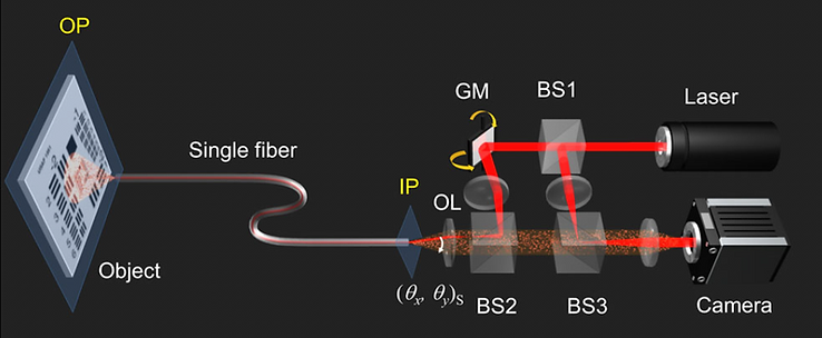
Recent trends in developing endoscopes is to gain the microscopic resolution and to reduce the diameter of the probes below a millimeter or so. The so-called endomicroscopes satisfying these two requirements provide a minimally invasive way of investigating the fine details of the microenvironments within the target organs. Typically, graded-index (GRIN) lens or image fiber bundles are widely used as imaging probes. In our studies, we used multimode fibers as imaging probes for further reducing the diameter of the unit. Since multimode fibers distort image information due to mode dispersion, bending and twist, we measured the transmission matrix of the fiber to recover the original image. In fact, our method enables us to use any light guiding media as an endoscopic probe. The examples of our investigation are given below.

Schematic layout of single-fiber microendoscope

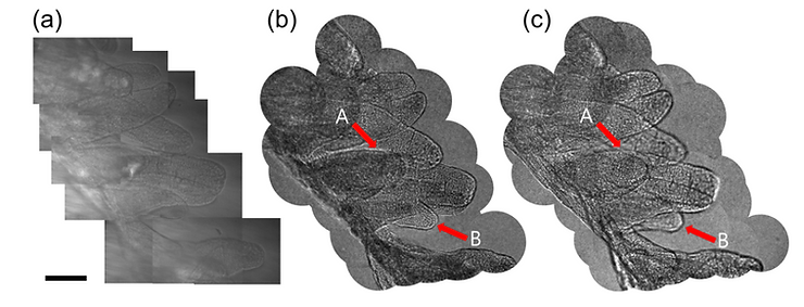
Examples of endoscopic imaging of rat villi. (a) Conventional transmission imaging. (b) Endoscopic imaging. (c) Numerical propagation of the image in (c)
References:
Youngwoon Choi, et al., "Scanner-free and wide-field endoscopic imaging by using a single multimode optical fiber," Physical Review Letters 109, 203901 (2012), Research Highlights in Nature 491, 641 (2012).
Changhyeong Yoon, et al., "Experimental measurement of the number of modes for a multimode optical fiber," Optics Letters 37, 4558 (2012)
Donggyu Kim, et al., Toward a miniature endomicroscope: pixelation-free and diffraction-limited imaging through a fiber bundle, Optics Letters 39, 1921 (2014)
Flexible, lensless, and label-free holographic endomicroscope
Ultrathin lensless fibre endoscopes offer minimally invasive investigation, but they mostly operate as a rigid type due to the need for prior calibration of afibre probe. Furthermore, most implementations work in fluorescence mode rather than label-free imaging mode, making them unsuitable for general medical diagnosis. Herein, we report a fully flexible ultrathin fibre endoscope taking 3D holographic images of unstained tissues with 0.85-μm spatial resolution. Using a bare fibre bundle as thin as 200-μm diameter, we design alensless Fourier holographic imaging configuration to selectively detect weakreflections from biological tissues, a critical step for label-free endoscopicreflectance imaging. A unique algorithm is developed for calibration-freeholographic image reconstruction, allowing us to image through a narrow andcurved passage regardless of fibre bending. We demonstrate endoscopicreflectance imaging of unstained rat intestine tissues that are completelyinvisible to conventional endoscopes. The proposed endoscope will expedite amore accurate and earlier diagnosis than before with minimal complications.

(a) Schematic of the experimental setup. (b) the image formation principle.

(a) Bright-field image of the fibre bundle The fibre cores where the illumination beam was focused are indicated as red dots. (b) Raw images captured by the camera (c) Complex field maps obtained from the raw interference images in (b). Circular colour map: real and imaginary values. (d) and (e) Fibre core-dependent phase retardations identified via the algorithm. (f) and (g) The same as (d) and (e), respectively, but for a low-contrast resolution target. (h) Conventional endoscopic image of a USAF target taken by the incoherent illumination from the IP. The fibreb undle was in contact with the target surface. (i) Coherent addition of inverse Fourier transformed images of the complex field maps in (c) before correcting for the fibre core-dependent phase retardations. (j) The same as (i) but after the correction.
References:
Choi, W. et al. Flexible-type ultrathin holographic endoscope for microscopic imaging of unstained biological tissues. Nature Communications 13, 4469 (2022).
Nguyen, T. V. A. et al. Object Function Retrieval by Model-Based Optimization in Fourier Holographic Endoscopy. ACS Photonics (2024) doi:10.1021/acsphotonics.3c01100.